When I first started working with 2D gels, streaking felt like my biggest enemy. I’d run what I thought was a perfect sample, only to see smears running horizontally or vertically across the gel. Instead of clean, crisp protein spots, I’d get hazy trails that hid the real story behind my sample. Over time, through trial, error, and plenty of frustration, I learned that streaking is not random—it’s the direct result of specific issues during sample prep, isoelectric focusing, equilibration, or SDS-PAGE. In this blog, I’m breaking down exactly what causes streaking in 2D gels and what I do to avoid it.
Understanding What Streaking Actually Means
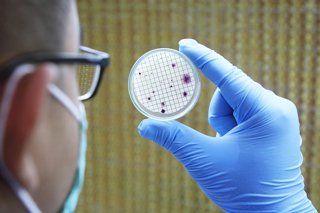
Before I go into the causes, I want to clarify what streaking represents. In my experience, streaking happens when proteins fail to focus into tight, distinct spots. Instead of resolving cleanly at their isoelectric point and molecular weight, they smear along the pH gradient (horizontal streaking) or along the SDS separation dimension (vertical streaking). Streaking tells me that something prevented the proteins from migrating properly.
The key is recognizing what went wrong—and that starts with understanding the root reasons behind streaking.
1. Inefficient Sample Solubilization
The first culprit I always check is solubilization. If proteins aren’t fully dissolved before loading onto the IPG strip, they clump, precipitate, and cause massive streaking. Hydrophobic proteins, membrane fractions, and samples with high lipid content are the most vulnerable.
I’ve learned that solubilization depends heavily on the right combination of chaotropes and detergents. Urea and thiourea help break apart protein structures, while detergents like CHAPS help keep them dissolved. When solubilization is incomplete, streaking becomes unavoidable.
I also test whether salts or nucleic acids contaminated the sample, because these can interfere with protein solubility. When I see strange horizontal smears, I always go back to my extraction strategy.
If you want to explore in more detail, click for more.
2. Salt Contamination or Improper Desalting
Salt is one of the biggest enemies of isoelectric focusing. Even a small amount can distort the electric field, cause current spikes, and push proteins into unwanted movements. Whenever I see inconsistent focusing or streaking across the entire pH range, salts are usually to blame.
I make sure I thoroughly dialyze, precipitate, or filter my samples before running 2D gels. For tricky samples—like cell lysates or bodily fluids—desalting becomes even more critical. High conductivity measurements before IEF often alert me to salt-related problems.
This is one of the issues I troubleshoot first because it’s extremely common and easy to overlook.
3. Overloaded Protein Samples
One thing I learned early on is that more protein does not equal better resolution. Overloading the gel causes proteins to bleed horizontally and merge into streaks. When I exceed the recommended micrograms for my IPG strip length, streaking becomes almost guaranteed.
If I start seeing streaks concentrated in high-abundance areas, I immediately check my protein load. For most samples, 50–100 µg works for analytical gels, and 100–500 µg works for preparative gels. Anything above that floods the focusing system.
I’ve found that sometimes adjusting sample load gives cleaner results than any other change I make.
4. Protease Activity Causing Protein Degradation
When proteases aren’t inhibited, proteins get partially digested during extraction and prep. Those fragments have different pI and MW values, which show up as streaks rather than clean spots. Whenever I forget to add protease inhibitors or don’t keep my sample cold, streaking becomes severe and unpredictable.
To prevent this, I process samples on ice and add a broad protease inhibitor cocktail immediately. Once degradation begins, the damage is irreversible.
5. Carbamylation Due to Urea Decomposition
Urea is essential in 2D electrophoresis, but it can betray you. When it decomposes—even slightly—it produces isocyanate, which reacts with proteins and shifts their pI. This chemical modification produces horizontal streaking because proteins no longer focus where they should.
I prevent this by using fresh, high-purity urea and never heating it above room temperature. I also avoid extended storage of urea-containing buffers. Carbamylation streaking is one of the most frustrating issues because it looks similar to other focusing errors.
6. Poor Rehydration of the IPG Strip
If the IPG strip is not fully and uniformly rehydrated, proteins do not distribute evenly across the pH gradient. Uneven rehydration leads to areas with too much salt, too little buffer, or inconsistent conductivity—all of which cause streaking.
I rehydrate strips overnight, use freshly prepared solutions, and seal them carefully to prevent evaporation. When strips dry out at the edges, I see localized streaks or distorted focusing.
This step seems simple but can make or break your 2D gel.
7. Improper Focusing Parameters
Even when the sample is perfect, poor IEF conditions can cause streaking. Voltage ramps, total volt-hours, and running time must be precise. If I rush IEF or skip steps, proteins won’t reach their isoelectric points.
Common mistakes I’ve made include:
- Running the system too fast in early steps
- Skipping low-voltage desalting periods
- Ending the run before the gel stabilizes
- Using incorrect pH strip ranges
Horizontal streaking is almost always related to focusing errors. I carefully document my voltage programs so I can easily troubleshoot.
8. Incomplete Equilibration Before SDS-PAGE
After IEF, proteins require proper equilibration in buffers containing SDS, DTT, and iodoacetamide. If this step is incomplete, proteins won’t bind SDS uniformly, leading to vertical streaking in the second dimension.
Issues arise when:
- I rush equilibration
- I don’t use enough buffer
- I fail to gently agitate strips
- Reagents like DTT lose strength
Equilibration prepares proteins for SDS-PAGE, so if it’s done poorly, the separation collapses.
If you’re curious about step-by-step buffer protocols, click for more.
9. Dirty or Oxidized Running Buffers
Contaminated buffers introduce artifacts that show up as background streaking or unusual smears. I’ve seen vertical streaks when SDS levels are too low or when buffers are reused too many times.
I always prepare fresh buffers for important runs and use molecular-biology grade reagents. When I started doing this consistently, I saw a dramatic reduction in streaking.
10. Sample Components That Interfere With Focusing
Some samples naturally contain materials that disrupt 2D gels:
- Lipids
- Polysaccharides
- Nucleic acids
- Phenolic compounds (common in plant extracts)
- Extremes of salt, detergents, or metabolites
These components interfere with protein solubility and focusing. I often treat samples with DNase, RNase, or precipitation methods to remove these contaminants. For high-lipid samples, additional detergents or organic extraction steps help.
11. Temperature Variations
Temperature shifts during IEF or SDS-PAGE destabilize gradients and can cause streaking. Even minor fluctuations can alter protein mobility.
I control temperature by:
- Keeping the IEF chamber cooled
- Using fresh buffers
- Preventing overheating during SDS-PAGE
Streaks caused by temperature are often faint but widespread.
12. IPG Strip pH Gradient Issues
If the pH gradient itself is defective or old, proteins won’t focus properly. Occasionally, manufacturing inconsistencies or improper storage lead to distorted gradients.
I always check expiration dates and store IPG strips at correct temperatures. Using low-quality strips has repeatedly given me poor results, so I don’t compromise here.
My Go-To Checklist to Prevent Streaking
To keep my gels clean and consistent, I follow this quick checklist:
- Fully solubilize proteins with the right detergents and chaotropes.
- Remove salts and interfering substances.
- Use correct protein load.
- Add protease inhibitors immediately.
- Use fresh urea and avoid heating.
- Rehydrate IPG strips overnight.
- Follow a proper voltage ramp for IEF.
- Equilibrate strips properly before SDS-PAGE.
- Prepare fresh buffers.
- Control temperature throughout separation.
- Choose high-quality IPG strips.
Every time I skip one of these steps, streaking inevitably shows up.
Why Professional Labs Help Reduce Streaking
Although I troubleshoot 2D gels myself, I know that experienced labs run them with far greater reproducibility. When I need publication-quality results, I turn to expert services like Kendrick Labs, Inc because they specialize in 2D electrophoresis and protein mapping at a level that’s difficult to replicate in-house.
Final Thoughts — You Can Eliminate Streaking With Consistent Technique
Streaking in 2D gels is frustrating, but it’s not mysterious. Each streak—horizontal or vertical—tells a story about what went wrong. The more attention I give to sample preparation, buffering, focusing conditions, and reagent quality, the cleaner my gels become. Over time, I’ve learned that consistency is everything.
If you ever need help interpreting gel behavior or troubleshooting your workflow, feel free to contact us anytime.